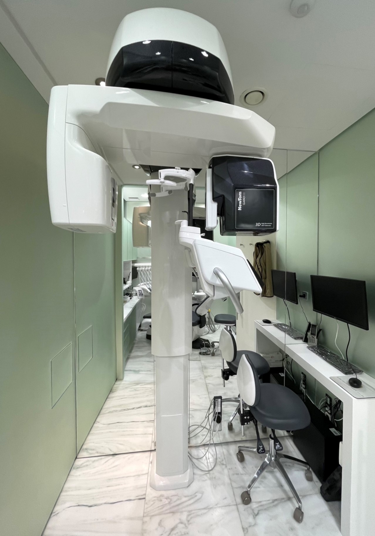
DIGITAL PANORAMIC RADIOGRAPH

DIGITAL PANORAMIC RADIOGRAPH
DIGITAL PANORAMIC RADIOGRAPH
X-rays are one of the most useful and widespread tools of the Medical science, as they can offer valuable information both for the initial diagnosis and for the planning and evaluation of the treatment course.
In order for the benefits to be maximized and for the negative effects of the radiation to the human health to be minimized, basic principles and rules of radiological protection have been established, that define the way radiation sources should be used.
In Greece, the competent body regarding the radiological protection of the population, employees and the environment, is the Greek Atomic Energy Commission (GAEC).
The Radiological Protection Regulations that are in force (Government Gazette no.216, Β, 6/3/2001), consist the basis of the radiological protection system in the country, as they describe the obligations and liabilities of all professionals that have to do with radiation, so that the use of radiation sources is safe and the lowest possible radiation impact is achieved. The Radiological Protection regulations are fully harmonized with the international and European regulatory framework.
After the radiograph, is there still radiation in the area?
No. X-rays are generated only when a radiograph takes place, which lasts only some fractions of a second. The percentage of radiation that is not absorbed by the imaging system is damped in the materials that surround the area. It is the same procedure as the generation of light by a common light bulb: when the circuit is interrupted there is no generation of light.
Do the materials that are irradiated by the X-ray tube become radioactive?
No, neither the materials that are irradiated nor the tube can become radioactive. The energy that is required in order for the materials to become radioactive is hundreds of times higher that the one which can be produced by a radiographic imaging device.
What measures of radiological protection must be used in Dental Offices?
In dental clinics X-ray (thyroid) collars and aprons should be available for radiological protection purposes, so that they are used in patients under 16 years old (collar) and patients that are in a reproductive age (apron). The use of such equipment by the dentists themselves is not obligatory, however it contributes to their better protection when they are in the radiography room.
How much safer is the position of the operator when he is covered by a wall or some other screening?
This depends on the material of the wall and its penetrability by radiation. The plastered brick walls practically eliminate the scattering radiation, while wood or plywood separators can be penetrated by X-rays. Lead (Pb) is the most effective screening material.
The operating license
As it is provided for in the Radiological Protection Regulations, for practices that include ionizing radiation a special license must be issued. In March 2008, Dr. Constantinos Laghios participated in the six month Oral Diagnostics and Radiology Training Program of the Dental School of the University of Athens with subject: Radiological Protection, Radiographic Technique, Radiodiagnosis.
Our Dental Clinic has a conformity certificate with No./408/1365/20.05.10


